


As a result of a person's upright walking, the spine, as an axial structure, took on the main load. That is why degenerative and dystrophic processes are quite common consequences of human life. One of the most common diseases of the musculoskeletal system is osteochondrosis, which brings great discomfort and can lead to disability. This article will discuss the most serious form of this pathology - common osteochondrosis.
general features
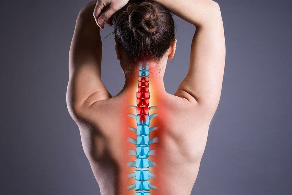
Osteochondrosis is a degenerative disease of the spine, which most frequently affects the thoracic, lumbar and cervical regions. This pathology is directly correlated with age. The disease is much more common in people over 40, but recently there has been a trend towards rejuvenation. Common osteochondrosis differs in that it affects more than one section of a department or several departments at the same time. Due to the progressive development of degenerative processes not only in the bone tissue, but also in the ligamentous apparatus of the spine, the vertebrae become mobile and exert pressure on the nerves and blood vessels. Symptoms of common osteochondrosis are associated with this, but it is worth noting that the disease can be asymptomatic for some time.
Important! The pathology requires multidisciplinary control, as it affects not only the musculoskeletal system, but also the nervous system, as well as internal organs. In addition to the spine itself, the pathological process can also affect other elements of the skeleton.
Etiology and pathogenesis
There are many reasons for generalized osteochondrosis. Some of them are associated with congenital skeletal defects, others with inadequate loading during vigorous activities. Particularly common factors that contribute to the development of the clinical picture are:
- injuries;
- flat foot;
- clubfoot - deformation of the foot (equinovarus, varus, valgus, depending on the position of the heel);
- work related to weight lifting;
- playing sports without warming up or warming up your muscles;
- work at low temperatures.
Low temperatures are considered provoking factors, as cold temporarily changes the molecular structure of soft tissues, reduces the intensity of blood circulation, reduces the conductivity of nerve impulses and metabolism, and therefore the functioning of the immune system. Other reasons disrupt the biomechanics of the spine and contribute to the rapid wear of intervertebral discs.
Pain in generalized osteochondrosis may be a consequence of osteophytes or disc deformation. The pain is usually radicular, that is, associated with compression of the posterior nerve roots.
Common osteochondrosis easily imitates other diseases. With injuries to the thoracic region, pain appears in the heart region and is confused with ischemic processes, and with injuries in the lumbar region - with radiculitis.
Symptoms
Clinical manifestations will depend on which parts are affected and in what combination.
When the cervical spine is affected, the following are characteristic:
- unstable blood pressure;
- headache;
- lack of coordination;
- hand pain;
- numbness in the upper body and arms.
For pathology in the thoracic region:
- intercostal neuralgia;
- stiffness in the arms and neck;
- dysfunction of internal organs.
If the lower back is affected:
- burn;
- urinary disorders;
- spasms;
- pain when walking.
Based on the above, it is easy to conclude that the pathology affects not only the spine and large joints, but also the autonomic nervous system. The latter is associated with disruptions in the functioning of internal organs. Common polysegmental osteochondrosis can sometimes get worse. In these cases, the manifestations are much more intense. With a combination of disorders from several departments, the symptoms will correspond.
Complications
Osteochondrosis can be conditionally divided into moderate osteochondrosis, which is a natural process of wear and tear on the spine as a result of vital activities, and severe osteochondrosis, which is more often characterized by complications.
Moderate osteochondrosis is easily treated with conservative treatment. And if it is impossible to completely stop the inevitable aging process, then it is quite possible to significantly slow it down. The complications that severe osteochondrosis can cause are as follows.
- Spondyloarthrosis.
- Intervertebral disc degeneration.
- Spinal stenosis.
Important! The intervertebral discs act as shock absorbers and reduce friction between the vertebrae. Degenerative processes in these structures can lead to protrusion of the nucleus pulposus of the disc and intervertebral hernia. Protrusion leads to compression of the roots and pain.
Spondyloarthrosis is the degeneration of the joints that connect adjacent vertebrae. Otherwise, these joints are called facet joints. When the articular cartilage is damaged, painful contact occurs between the vertebrae. With degeneration of the facet joints, bone growths appear more often, which leads to spondylosis.
Stenosis is a narrowing (in this case, of the spinal canal). Typically, stenosis is the result of pathologies such as intervertebral hernia or spondylosis. Bone growths and hernial protrusions compress nerve roots at their entry and exit points.
The clinical picture of severe osteochondrosis is the result of complications:
- chronic back pain;
- friction of bone surfaces;
- stiffness;
- sudden muscle weakness;
- decreased reflexes;
- tingling in the limbs;
- radiating pain;
- sciatica symptoms.
Sciatica is caused by compression of the sciatic nerve.
Classification
There are four degrees of osteochondrosis. Classification occurs on the basis of the collected history and with the help of instrumental diagnostic methods. The main criteria for this classification are pain and neurological symptoms.
- Grade I - pain is easily relieved with medication.
- Grade II - characterized by prolonged pain and deformation of the spine with moderate neurological symptoms.
- Grade III - pain is systematic, neurological symptoms are significant.
- Grade IV - constant pain, multiple neurological deficits. Disturbance in the conduction of nerve impulses. Paralysis and paresis.
In the case of generalized dysplastic osteochondrosis, the patient is assigned disability status. Depending on the general condition of the patient, the degree and intensity of the development of the clinical picture, the disability can be of three groups.
Types of disability in osteochondrosis.
| Group | Description |
|---|---|
| First group | Spine functions are lost. The patient is unable to move independently and take care of himself. |
| Second group | The patient is able to move and perform small tasks, but periods of exacerbation are frequent. The operation is contraindicated or useless for some reason. Or surgery has already been performed, but turned out to be ineffective. |
| Third group | The patient is able to take care of himself. There is pain and vestibular symptoms, but the frequency of exacerbations is moderate and periodic. |
The disability group is assigned by the doctor based on some studies to assess work ability.
Diagnosis
When visiting a doctor, the diagnosis will consist of several components. The first and most important is the collection of anamnesis based on subjective information provided by the patient. Attention is paid to family history, as osteochondrosis has a genetic component. The specialist asks about the place of work, living conditions and the course of the disease itself, and the patient must describe exactly what bothers him. The best results can be achieved with good feedback between the patient and the doctor.
The next method is an objective study, carried out by the specialist himself or using instrumental methods. The doctor checks the range of motion in the neck and limbs, which may be visibly reduced due to pain and stiffness. Using the palpation method, he records how much the muscles are spasming and how curved the spine is. Attention is drawn to a neurological examination, with which weakened reflexes can be tracked. This symptom may be the result of compression or damage to the nerve.
Instrumental methods for diagnosing common osteochondrosis include:
- Radiograph of the entire spine in two projections.
- Magnetic resonance imaging to evaluate ligaments and nervous tissue.
- An electrophysiological study to test the conduction of nerve impulses.
Radiography is effective in determining the presence of bone growths - osteophytes, narrowing of the spinal canal and the presence of other diseases arising from osteochondrosis, for example, scoliosis.
Computed tomography can also be used in conjunction with MRI. Using a CT scan, you can determine the degree of compression of the nerves by the spurs.
The diagnosis of generalized polysegmental osteochondrosis is made if other pathologies causing destruction of the vertebrae (for example, tuberculosis) are excluded and if several segments of one or more departments are affected.
There are additional diagnostic methods. These include:
- Bone scan.
- Discography.
- Myelogram.
A bone scan can detect conditions such as osteoarthritis, fractures, or infections. This method is radionuclide and is suitable for differential diagnosis and determination of possible complications.
During discography, a contrast agent is injected into the nucleus pulposus of the intervertebral disc. This method is effective in determining the presence of a herniated disc.
Myelogram is also a contrast research method. Contrast is injected into the spinal canal and the image is recorded using x-rays or computed tomography. Using this method, you can determine the condition of the spinal cord, the presence of narrowings and compressions.
Treatment
Treatment is based on the following mechanisms.
- Slow down the degenerative process, improving the supply of nutrients to the structures of the musculoskeletal system.
- Spinal stabilization.
- Elimination of compressed nerve fibers.
- Relieving symptoms.
The following medications are used for drug treatment:
- nonsteroidal anti-inflammatory drugs that relieve inflammation and pain;
- Anilides relieve pain in the early stages;
- local analgesics in the form of ointments;
- muscle relaxants to reduce muscle spasms and increase range of motion;
- B vitamins to improve the conductivity of nervous tissue;
- chondroprotectors, which reduce the rate of progression of degenerative processes through the integration of active substances (condotin sulfate and glucosamine) into cartilage cells. As a result, metabolism is normalized and clinical manifestations are reduced. The medicine has been used for a long time and requires special consultation during pregnancy, lactation and the presence of gastrointestinal diseases. An absolute contraindication is phenylketonuria;
- antispasmodics relieve spasms of smooth muscles and thereby alleviate the manifestations of osteochondrosis in internal organs;
- antioxidants;
- antidepressants to eliminate the psychosomatic component of this disease. They interfere with the transmission of nerve impulses from the central nervous system to the brain. Promotes the production of endorphins and helps solve the problem of chronic insomnia due to constant pain.
- neuropathic agents to eliminate damage to nerve endings.
- opiates for unbearable pain and ineffectiveness of other analgesics.
The following are used as invasive medical procedures:
- steroid injection into the epidural space. Steroids are powerful anti-inflammatory medications. They relieve inflammation of the nerve roots, which helps relieve pain caused by radiculopathy. Complicated procedure. Requires a qualified specialist;
- facet joint injections. Injected drugs cause local numbness and pain relief.
It's important to know! Taking medications is not intended to eliminate the disease - there are no medications that can completely eliminate osteochondrosis, which is a chronic disease. Medications are prescribed only to alleviate symptoms.
Medications are prescribed by the attending physician. The patient is informed about the possible side effects of each medication and then decides for himself which treatment to choose.
For symptoms that give reason to suspect osteochondrosis, contact a vertebrologist, orthopedist and neurologist. High-quality medical care will consist of the close cooperation of these specialists with each other and with the patient.
Physiotherapy
Physiotherapy is used as a set of auxiliary therapeutic measures to improve blood circulation and metabolism in the affected tissue. For generalized osteochondrosis, the following methods are used.
- Electrophoresis (based on the movement of colloidal particles under the influence of an external electric field).
- Phonophoresis (a combination of ultrasound and medication).
- Magnetotherapy (use of static magnetic field).
- UHF therapy (ultra high frequency therapy).
- Electromyostimulation (stimulation of nerves and muscles).
- Acupuncture (acupuncture).
- Laser exposure.
In addition to physiotherapy, manual therapy and physiotherapy are actively used. Professional massage can lead to long-term remission. Therapeutic exercises should not be performed during an exacerbation of the disease, as they can lead to complications. During the period of remission, moderate physical activity maintains muscle tone and, therefore, the tone of the spine. Exercises are performed under the supervision of an instructor and prescribed by the attending physician.
During an exacerbation, you cannot warm the spine, but you can wear a corset, but only for a few hours. In other cases, wearing a corset for more than a few hours is not ideal because it can lead to muscle atrophy.
Manual therapy can increase clearance of pinched nerves and reduce neurological symptoms. Alternative methods are leeches and vacuum massage. These methods aim to improve blood circulation in the affected area. Sanitary spa treatment is useful. Particular preference is given to water procedures.
Surgery
When treating osteochondrosis, specialists are more willing to resort to conservative therapy, however, for proper effect, a lot of time, patience and scrupulous adherence to the patient's recommendations are required. If conservative treatment is ineffective, only then should you resort to invasive methods. Generally the operation is palliative. This means that the operation will be performed only to alleviate symptoms and comparatively improve the quality of life, but not for complete cure (it is fair to say that conservative treatment also does not lead to complete elimination of the disease, but rather to the patient's receptivity to non-invasive therapy is a sign of a good prognosis).
There are two types of surgery: decompression and stabilization. The first aims to relieve nerve compression and the second aims to stabilize the spine. The following operations are classified as decompression operations.
- Facetectomy – removal of joints to relieve compression.
- Foraminotomy is an increase in the lumen of the spinal canal, which has narrowed due to osteophytes.
- Laminectomy is the removal of the back part of the vertebra, which may be deformed due to osteochondrosis.
- Laminotomy - removal of a fragment of the back of the vertebra to widen the spinal canal.
These operations require a posterior approach, but in the case of an intervertebral hernia the surgical approach will be anterior.
Decompression surgeries with anterior approach are as follows.
- Discectomy – removal of an intervertebral disc.
- Corpectomy – removal of the entire vertebral body with adjacent discs.
Stabilization operations include:
- Spinal fusion is a method of fusing vertebrae.
- Artificial intervertebral disc.
The need for stabilizing operations arises after discectomy.
Surgeries are rarely prescribed because there is a risk of developing serious complications.
Complications include:
- pain relapses;
- false union;
- infection;
- phlebitis in the limbs;
- violation of the act of urination;
- pain due to the graft;
- failure of built-in fasteners.
The postoperative period lasts several months. The sutures heal 2 weeks after surgery. If complications are detected, you should immediately consult a doctor.
After the operation, a rehabilitation course is carried out to speed up the healing process and restore full working capacity.
General recommendations
Proper nutrition helps prevent relapses of exacerbation of common osteochondrosis. Proper nutrition is primarily necessary to maintain a stable body weight, as excess weight puts additional pressure on the spine. In this case, the diet must be complete, fortified and rich in calcium, magnesium and potassium. It is necessary to limit coffee consumption as it eliminates calcium from the body. It is useful to visit the pool. You should avoid staying in the same position all the time.
If there are frequent exacerbations and lack of discipline to strictly follow the recommendations, it is best to undergo complete treatment in a hospital under the supervision of a doctor.
You cannot take medication alone.
Let's summarize
Often, generalized osteochondrosis develops after an incompletely cured "single" osteochondrosis. This fact suggests that if any discomfort occurs, you should immediately seek medical attention, without waiting for the pain to disappear on its own. In this case, it will be much easier to prevent the development of other pathologies and even more so to cure the root cause.